

This post will introduce a few abnormal findings in chest x-rays including pneumonia, abscess, cardiomegaly, and medical devices. There is great interest in building machine learning algorithms to automatically detect abnormal findings in chest x-rays. For a review of normal chest x-rays, see this post. For a review of basic chest anatomy, see this post.
Pneumonia & Abscess

The above image (source) shows a patient with pneumonia and abscesses. Recall that the lungs are supposed to be dark, but we can see cloudy grey inside the lungs, particularly on the left side of the patient (the right side of the picture, with the “L” in the upper corner.) The cloudy grey represents infected lung tissue, i.e. the pneumonia. You can also see a darkened circle, which represents an abscess:

Cardiomegaly
“Cardiomegaly” is an enlarged heart (from Greek “cardio”=heart and “megas”=great). An enlarged heart can occur for a variety of reasons including high blood pressure, heart failure, cardiomyopathy (a disease of the heart muscle), thyroid disease, and viral infection of the heart.
Cardiomegaly can be determined by measuring the “cardiothoracic ratio“:

The cardiothoracic ratio is calculated as (MRD+MLD)/ID where MRD is the greatest perpendicular diameter from the midline (where the spine is) to the right heart border, and the MLD is the greatest perpendicular diameter from the midline to the left heart border. The ID is the internal diameter of the chest at the level of the right half of the diaphragm. The cardiothoracic ratio should be less than 0.5.
Here is a chest x-ray from someone with an enlarged heart. You an also see a pacemaker in the image:
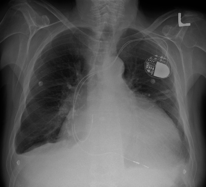
Additional examples: Normal heart, Enlarged heart
Devices
Here’s another chest x-ray showing a medical device:

Chest X-Ray with ICD. In this image, you can see an ICD, or “implantable cardioverter defibrillator.” An ICD is an extremely useful medical device that can prevent sudden death in patients who have problems with their heart rhythm. The device is implanted inside the body and tracks the heart rate. Wires (labeled “pacing lead” and “shock lead”) connect the ICD generator to the heart. If the device detects a dangerous heart rhythm, it will shock the heart to restore a normal heartbeat. If you have ever watched a medical TV drama, an ICD is like a miniaturized, implanted version of those paddles that they put on the chest and yell “CLEAR” to wake someone up.
A note about the featured image
The featured image for this post is a blue-tinged chest x-ray. This is actually a completely normal chest x-ray; the digitization was just performed in an unusual manner. A patient obtained a copy of their chest x-ray film and hung it over their computer screen, then took a digital photo (hence the weird blue glow.)
Stay tuned – in a future post I’ll go over a few more interesting findings in chest x-rays!
Want to be the first to hear about my articles bridging healthcare, artificial intelligence, and business—and get a free list of my favorite health AI resources? Sign up here.

Comments are closed.