

Today we’ll talk about additional interesting chest x-ray findings, including nodules, masses, atelectasis, scarring, pneumothorax, pleural effusion, rib fractures, and heart failure.
Relevant review posts: normal chest anatomy, normal chest x-rays, abnormal chest x-rays part I.
Nodules, Masses, and Tumors
Quick summary of terminology:
- a nodule is smaller than 3 cm in diameter
- a mass is bigger than 3 cm in diameter
- a “tumor” is some kind of abnormal tissue growth. It can be non-cancerous (benign) or cancerous (malignant)
- Thus, a small tumor (<3 cm) could show up as a “nodule” on an x-ray, and a big tumor (>3 cm) could show up as a “mass.”
The above image shows a “solitary pulmonary nodule“, also known as a “coin lesion” because it’s roughly coin-shaped and coin-sized. 80% of coin lesions are benign, and 20% are malignant.
Note that an actual coin (like a penny or a quarter) would show up as super white – e.g. this x-ray of a swallowed coin – because coins are metal and reflect x-rays.
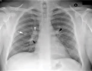
Not all nodules are “solitary pulmonary nodules.” There might be multiple nodules.This image shows multiple lung nodules (indicated by the black and white arrows), which were caused by tuberculosis infection:
Additional examples of chest x-rays showing nodules or masses:
- Large mass lesion
- Tumor in the left lung
- Tumor in the right lung
- Small cancer in right upper lung
- Cancer tumor colorized pink
This article, “Evaluation of the Solitary Pulmonary Nodule” discusses different causes of solitary pulmonary nodules, and next steps for evaluation.
This article, “Differential diagnosis and evaluation of multiple pulmonary nodules” discusses different causes of multiple pulmonary nodules.
Atelectasis vs. Scarring
Atelectasis and scarring are two completely different things, but they look quite similar on a chest x-ray.
- Atelectasis is collapse of lung tissue or a failure of lung tissue to expand – i.e., instead of being light and full of air, the lung tissue has had the air squished out of it. Because the collapsed tissue is more dense, it appears whiter than the surrounding normal lung. (Recall that the lungs are supposed to be black on a chest x-ray.)
- Scarring in the lung is similar to scarring on the skin – it’s a result of damage to the lung (e.g. from a severe infection.)
This page and this page show some examples of atelectasis.
Here’s a really severe example of scarring in the lung (and a metal necklace):

In this case, the diagnosis is “pulmonary fibrosis” which is just the medical term for “scarring of the lungs.” This particular patient was believed to suffer pulmonary fibrosis due to a medication complication.
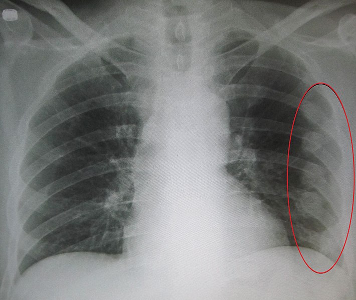
Pneumothorax
All of the air in your lungs is supposed to be contained within the spongy tissue of the lung. If air leaks out of the lungs and begins to fill up the space between the lungs and the inside of the chest, it’s referred to as a “pneumothorax,” shown in this image:
The chest on the left side of the image looks much darker than the the right side because it’s full of pure air, instead of lung tissue plus air. Here I’ve outlined the collapsed lung in red, and labeled the free air:
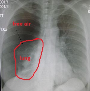
This is very bad because the collapsed lung isn’t functioning at all — it’s not delivering any oxygen to the body. Luckily, there is still one functioning lung, but it’s important to fix the collapsed lung and help it inflate again.
But wait…I thought atelectasis was a collapsed lung!
- Atelectasis refers to the actual part of the lung tissue that is collapsed, while pneumothorax refers to air that is present inside the chest cavity outside of the lung tissue. It’s possible to have atelectasis without pneumothorax (e.g. you’d have atelectasis only if a small part of the lung has collapsed without any free air in the chest cavity.)
- Atelectasis can be seen after chest surgery due to collapse of the alveoli (the small air pockets in your lungs). Atelectasis can also be caused by pneumothorax (i.e. by free air getting in to the chest space.)
- Pneumothoraces can happen spontaneously (e.g. in tall thin people, a “bleb” in the lungs can rupture), or they can be caused by injuries (e.g. puncture of the chest wall by a knife or a fractured rib) or illness (damage to the lungs from pneumonia, tuberculosis, etc.)
Pleural effusion
Pleural effusion refers to excess liquid surrounding the lungs. There are different names for pleural effusions depending on what kind of liquid is surrounding the lungs: hydrothorax (for a water-like fluid), hemothorax (blood), pyothorax or empyema (pus), and urinothorax (urine – yes, it’s possible, but it’s rare).
Here’s a chest x-ray showing a huge pleural effusion:

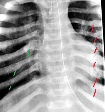
Rib fractures
This image shows fractured ribs, circled in red.
Here’s another example of multiple fractured ribs, with the fractures indicated via arrows:
Heart failure
Here’s an example of a chest x-ray showing intermediate severity heart failure:

A chest x-ray showing heart failure encompasses a few different concepts we have already covered, including cardiomegaly (increased heart size/increased cardiothoracic ratio) and pleural effusion. Two additional chest x-ray signs of heart failure are:
- “Kerley B lines” (not to be confused with Cardi B…): short parallel lines at the edge of the lungs
- “cephalization of vessels“: the blood vessels in the upper chest (closer to the “cepha” or head) are more prominent due to high blood pressure in the pulmonary veins
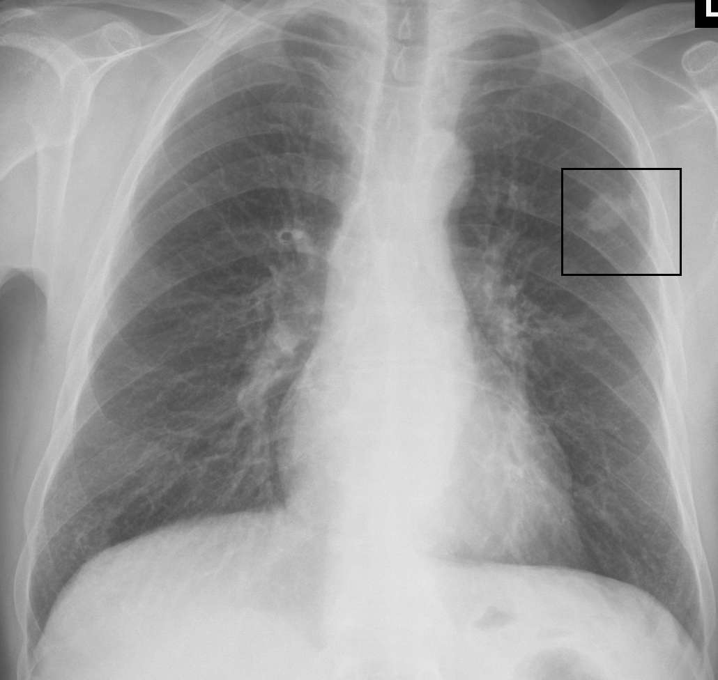
Extra image
This extra image is an abnormal chest x-ray demonstrating a finding from Abnormal Chest X-Rays Part I. Can you guess what it is?
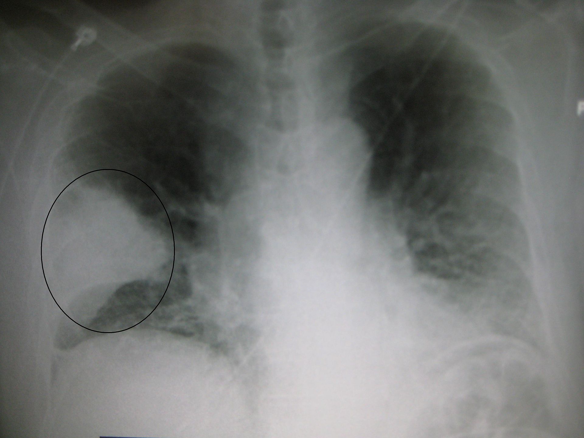
It’s pneumonia! The circle surrounds a wedge-shaped area of airspace consolidation indicative of acute bacterial lobar pneumonia (i.e., the circled lobe of the lung has been infected with bacteria.)
Stay tuned for future posts on more chest x-ray abnormalities, as well as an overview of how machine learning can be used to automatically identify abnormalities in chest x-rays.
As an independent researcher (MD + AI PhD + 7 yrs prior founder/CEO experience), I build and evaluate cutting-edge healthcare AI for startups. Contact me to learn more.
Want to be the first to hear about my articles bridging healthcare, artificial intelligence, and business—and get a free list of my favorite health AI resources? Sign up here.

Comments are closed.