

This post will wrap up abnormal findings in chest x-rays, with an overview of pericardial effusion, hiatal hernia, pneumonectomy, COPD, artificial heart valves, breast implants, and tubes!
Pericardial effusion
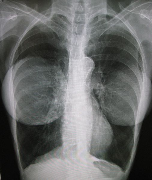
The above chest x-ray shows a huge pericardial effusion. A pericardial effusion is a condition where the sac surrounding the heart (the pericardium) fills up with fluid, like a water balloon. The heart can actually swing around inside of this huge sac of fluid, as shown in this gif (which was created using ultrasound, not x-ray):

Pericardial effusions can be caused by infections (e.g. tuberculosis or coxsackie virus), certain drugs, and inflammatory disorders like lupus.
If a pericardial effusion becomes too severe, it can actually prevent the heart from pumping blood due to the extremely high pressures of the fluid around the heart. This is dangerous and has to be treated immediately by draining away the fluid.
Additional x-ray images of pericardial effusion are available here.
Hiatal hernia
In a hiatal hernia, a hole in the diaphragm allows the stomach to squeeze into the chest cavity. The image below shows a severe hiatal hernia (source). The the herniated abdominal contents have been circled in green, and the heart has been circled in red:


Click here for another example of a hiatal hernia chest x-ray.
Pneumonectomy
A pneumonectomy is the surgical removal of a lung. The most common reason to remove a lung is lung cancer.
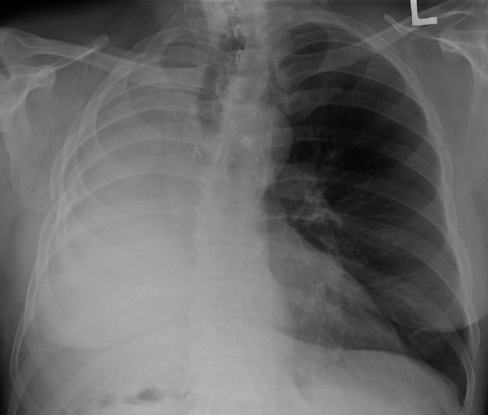
In the below chest x-ray, the right lung has been removed. The half of the chest that looks white instead of black is the missing lung. The remaining lung appears black, and is on the side marked with the “L.”
Immediately after a pneumonectomy, the empty space fills up with air. Then, over several weeks to months, the empty space fills up with fluid (reference).
Chronic obstructive pulmonary disease
You may have heard of chronic obstructive pulmonary disease (COPD) referred to by the older terms “emphysema” (indicating particular kinds of lung changes) or “chronic bronchitis” (indicating particular symptoms including coughing up phlegm).
COPD is a lung disease most commonly caused by tobacco smoking or air pollution. In rare cases, a patient may suffer from COPD due to a genetic disorder.
In COPD, the normally spongy lungs become filled with large holes, or “bullae,” which makes the lungs appear even darker on a chest x-ray. Here’s an example of lung bulla (a hole or “blister”) circled in black, from a patient who has COPD due to the genetic disorder alpha-1 antitrypsin deficiency:

Artificial heart valves
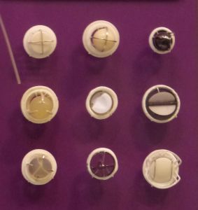
Sometimes, the natural heart valves stop working (e.g. due to infection). This makes it difficult for the heart to pump blood properly. In order to prevent damage to the heart, the malfunctioning valve(s) must be replaced. A variety of artificial heart valves are available, including the designs shown in the image below:
Artificial heart valves show up in chest x-rays as white rings. Here’s a chest x-ray showing four artificial heart valves at the same time. Here’s an image showing two.
Breast implants
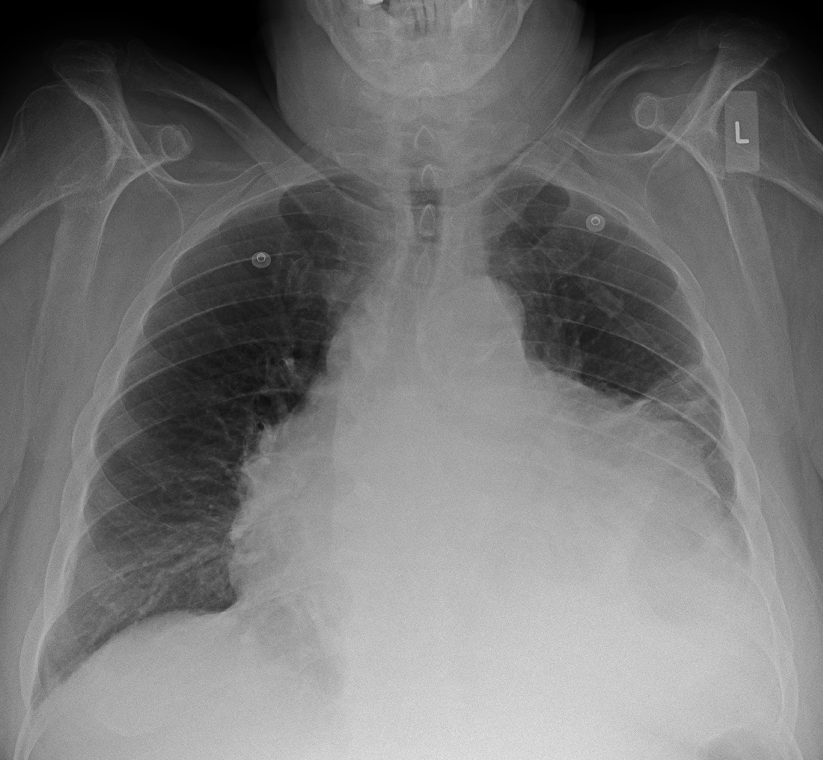
Here’s a chest x-ray showing bilateral breast implants. Because the implants are denser than normal tissue, they show up as lighter in color:
Tubes
A patient may need a tube inserted for a variety of reasons. Chest x-rays can be used to double check that the tube has been positioned appropriately.
- Nasogastric tubes go from the nose to the stomach, and can be used to feed a patient.
- Endotracheal tubes go from the mouth into the trachea (the windpipe), and are used for ventilation (breathing machines.)
- Tracheostomy tubes are inserted directly into the windpipe through a hole made in the neck. They can be used to help with breathing when an endotracheal tube is not an option (e.g. when a breathing machine will be required for longer than a couple weeks)
- Pleural tubes or “drainage tubes” are inserted directly into the chest cavity through the skin and muscles, for example to help drain a pleural effusion
“A pictoral essay: Radiology of lines and tubes in the intensive care unit” includes example x-rays for all of the aforementioned tubes.
Interrupted chest x-ray
I’m not sure who took this picture, but they weren’t following standard protocols…
Additional Links
- This image shows BB gun pellets (white dots) after an unfortunate BB gun accident
- This image shows a metallic foreign body stuck in the upper esophagus
- This image shows a left ventricular assist device (LVAD, pronounced “ELL-vadd”) which is a type of pump that can be used to assist a failing heart
- Radiography of Cardiac Conduction Devices: many different x-ray images of pacemakers
- Medical Devices of the Chest: scroll to the end for tons of example chest x-rays showing catheters, tubes, wires, and heart valves
Featured Image
What’s going on with the featured image??

This x-ray has been inverted and “enhanced” so that dark appears light and light appears dark. This wouldn’t be used in a medical setting but it does look interesting!
Finally…
This “x-ray video” was created using projectional renderings of a CT scan:

Thanks for reading! For more examples of abnormal chest x-rays, check out Part I and Part II of this series.
Want to be the first to hear about my articles bridging healthcare, artificial intelligence, and business—and get a free list of my favorite health AI resources? Sign up here.

.gif)
Comments are closed.