

X-rays are the oldest and most popular form of radiology imaging, and automated interpretation of chest x-rays is a a fun machine learning task. In this post, I’ll introduce the basics of chest x-ray interpretation.
Every year medical personnel perform 3.6 billion medical procedures involving ionizing radiation. A large number of these procedures are projectional x-rays (commonly just called “x-rays”), where a very small dose of radiation is used to create a 2-D pictures of the chest, abdomen, pelvis, or limbs.
Chest x-rays in particular can be used to identify a variety of findings, including pneumonia, pneumothorax (air pockets inside the chest), heart failure, rib fractures, lung masses, big lymph nodes, effusions (extra fluid in the chest), and hernias through the diaphragm, among other conditions. But in order to understand what these abnormal findings look like on a chest x-ray, it’s important to first learn what a healthy chest x-ray looks like.
Check out “Anatomy for Radiology: Chest” for a review of basic chest anatomy.
Chest X-Ray Coloration
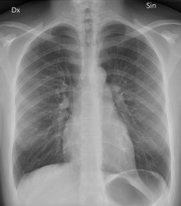
Normal Chest X-Ray. This image shows a normal chest. Darker colors indicate less dense material, and lighter colors indicate more dense material. The lungs appear black because they are spongy with a lot of air content. The bones appear white because they are hard, mineralized, and block x-rays effectively.
The Ribs
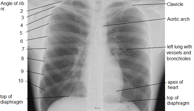
Chest X-Ray: Ribs. In the figure above, the ribs have been numbered 2 through 10. There are actually 12 ribs on each side; ribs 1, 11, and 12 are not numbered here. The ribs look like light grey stripes. You can also see the clavicle, a v-shaped bone across the top (aka the “collarbone”. In birds, the collarbones are fused to form the “wishbone”).
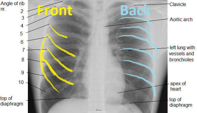
Anterior vs. Posterior Ribs. Here I’ve added yellow and blue lines to show the difference between the anterior sections of the ribs (front, in yellow) and posterior sections of the ribs (back, in blue). Recall that the ribs wrap all the way around the front and the back of the chest to protect the lungs and heart. On a chest x-ray, you can see both the front sections and the back sections of the ribs, just like the shadow of a cage projected onto a wall.
Here, the anterior ribs are underlined in yellow, and point more diagonally downwards. If you feel your own rib cage in the front, you can recognize the downward-pointing angle. The posterior ribs are underlined in light blue, and look more horizontal.
See if you can identify the back ribs on the side where the front ribs are highlighted, and if you can identify the front ribs on the side where the back ribs are highlighted.
Heart, Great Vessels, etc.

Here, I’ve outlined a few more structures:
- Heart and Great Vessels: The heart and some of the great vessels (including the aorta) are outlined in red. Recall that in most people the heart points down and to the left, which you can see here. The left side of the patient is the right side of the picture.
- Diaphragm: The diaphragm is the thin dome-shaped muscle that helps with breathing (and hiccups). It is outlined in orange. It separates the chest from the abdomen.
- Liver: The liver isn’t outlined, but its position is indicated by the purple label. The liver is on the right of the patient (left of the picture) and looks like smooth grey on a normal chest x-ray.
- Stomach: The stomach isn’t outlined, but its position is indicated by the green label. Air in the stomach is normal, and is referred to as the “gastric bubble.”
- Clavicle: I’ve underlined the clavicle (collarbone) in blue.
Lungs
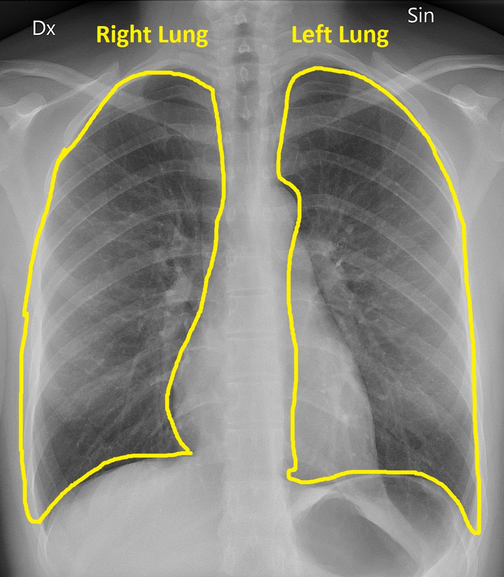
Here I’ve outlined the lungs in yellow. You can see that part of the left lung overlaps the heart. The lungs are the darkest part of the chest x-ray because they are the least dense.
Side note on…Radiation!
A chest x-ray results in an effective radiation dose of 0.1 mSv, which is the equivalent of natural background radiation for 10 days, or about three flights from the East Coast to the West Coast of the United States (each flight results in 0.03 mSv of radiation exposure, because being at higher altitude results in less protection from cosmic rays). Radon gas in houses over one year results in 20x more radiation exposure than a chest x-ray. Overall, a chest x-ray is a very small amount of radiation for a very useful test! (Radiation Doses Reference)
Just for Fun
Here are some neat animal x-rays: a chameleon, flying fox, ball python, beaver, and toucan, from the Oregon Zoo.
Aaand…some x-rays of flowers from the 1930s.
More Annotated (Human) Chest X-Rays:
- Life In The Fast Lane Normal Chest X-Ray
- Components of the Heart and Great Vessels (scroll down to second image)
- Components of the Lung
The end! 🙂 In a follow-up post, I’ll introduce chest x-ray pathology: i.e., the visual findings that we want to teach machine learning algorithms to detect.
As an independent researcher (MD + AI PhD + 7 yrs prior founder/CEO experience), I build and evaluate cutting-edge healthcare AI for startups. Contact me to learn more.
Want to be the first to hear about my articles bridging healthcare, artificial intelligence, and business—and get a free list of my favorite health AI resources? Sign up here.

_chest_radiograph_(X-ray).jpg)
Comments are closed.