

In this post, I will introduce the anatomy of the abdomen. Abdominal x-rays and abdominal CTs are common imaging studies performed in a variety of medical settings. First I will introduce the concepts, organs, and structures related to the abdomen, and then I will go over some diagrams.
Path of Food
One popular organ in the abdomen is the stomach, but the stomach is just one stop in the journey of food. It can be helpful to understand the path of food through the body in order to understand the abdomen (which contains most of that path.) Voila:
- Mouth. The mouth starts the digestive process by mixing food with saliva. The tongue can taste at least five flavors: sour, sweet, salty, bitter, and umami (triggered by monosodium glutamate, aka MSG.) Recent evidence suggests that there may be additional fundamental flavors: fat and “complex carbohydrate” (e.g. bread, rice). The finer details of different flavors actually come from the nose (i.e. smell.)
- Esophagus. The esophagus is the tube that carries food from your mouth, through your chest, and into your stomach. In its resting state, the esophagus resembles a flattened soft pink hose – it’s not propped open. This is contrast to the windpipe, which is a hollow tube held open at all times by the firmness of its walls. The esophagus runs behind the windpipe and in front of the spine.
- Stomach. Your stomach can hold about a half-pound of food. It mixes food with hydrochloric acid and digestive enzymes. Some cells in your stomach called “parietal cells” are responsible for making the hydrochloric acid.
- Small intestine. Many people think that the stomach extracts energy from food, but it is actually the small intestine that extracts nutrients from food and pumps these nutrients into the blood.
- Duodenum: the first part of the small intestine. The name “duodenum” comes from the Latin word “duodeni” meaning “in twelves” because the length of the duodenum is approximately the breadth of twelve fingers.
- Jejunum: the middle part of the small intestine. The name “jejunum” comes from the Latin word “jejunus” meaning “fasting”, because the jejunum is often found empty after death.
- Ileum: the last part of the small intestine. The name “ileum” (or “ilium”) comes from the Latin word “ilia” meaning “flanks, entrails.”
- Large intestine. The large intestine extracts water from food and pumps the water into the blood. The large intestine is divided into five sections: the ascending colon (food moves up), the transverse colon (food moves across), the descending colon and sigmoid colon (food moves down), and the rectum (food moves out).
Additional Organs and Structures of the Abdomen
- The appendix. The appendix is a small finger-like sac that protrudes from the ascending colon. There are many theories about the purpose of the appendix, including (a) the appendix helps maintain gut bacteria by protecting them in case of serious illness, and (b) the appendix serves as part of the immune system. Recent evidence suggests that people who have had their appendix removed have a lower risk of Parkinson’s disease but a higher risk of certain autoimmune conditions like lupus and rheumatoid arthritis.
- The gallbladder. This is a hollow organ (sac) that stores bile. The gallbladder squirts bile into the small intestine, where the bile helps with fat digestion.
- The liver. This is a solid organ that detoxifies the blood. For example, the liver detoxifies alcohol by turning it into acetate. Genetic variation in the alcohol detoxification pathway causes certain people to be more prone to alcohol flush reactions. The liver also produces bile, which is then sent to the gallbladder for storage.
- The spleen. This is a solid organ that stores white blood cells for the immune system and helps recycle red blood cells. About 10% of people have extra, miniature spleens called “splenules.”
- Pancreas. This is a squishy organ that produces digestive enzymes and produces insulin to help the body absorb sugar. If a patient’s pancreas cannot produce insulin, then that person has diabetes.
- Kidney. There are two kidneys, one on the right and one on the left. The kidneys are solid organs shaped like kidney beans (or rather, kidney beans are shaped like kidneys…) The kidneys filter wastes (e.g. urea) out of the blood, and produce urine that contains these wastes. If a patient’s kidneys fail then that patient must undergo dialysis to survive. A dialysis machine is basically a giant artificial kidney.
Adjectives
- “Renal” refers to the kidneys
- “Helaptic” refers to the liver
- “Splenic” refers to the spleen
- “Colonic” refers to the colon
Pictures of the Abdomen
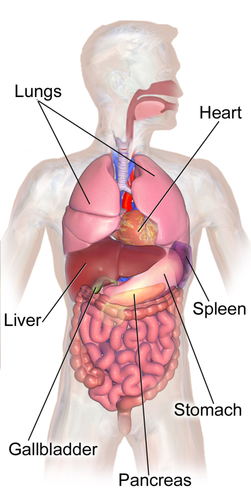
Various Abdominal Organs (Image Source). Here you can see the liver, which sits on the right side of the body (the left side of the picture). The stomach and spleen are on the left side of the body (the right side of the picture.) The pancreas sits behind the stomach. The large and small intestines are also visible but not labeled here.
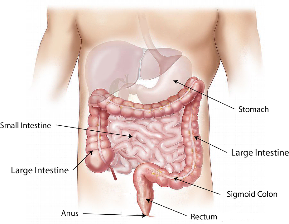
The Intestines (Image Source). Here you can see how the large intestine (shown here in darker pink) forms a kind of frame around the small intestine (shown here in lighter pink). Food travels first through the small intestine, which really does look “disorganized” in real life, and then through the large intestine, which is “pinned” against the “back wall of the body” so that it hangs in that frame-like shape.
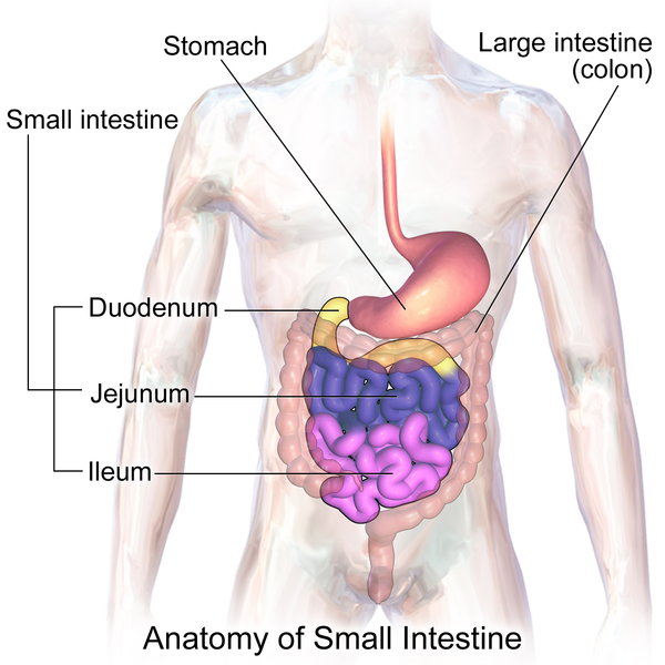
The Small Intestine (Image Source). Here, the difference pieces of the small intestine have been colored in different colors. The duodenum is the first piece of the small intestine, which is shown in yellow and is attached to the stomach. The jejunum is the second piece, shown in dark blue. The ileum is the final piece, shown in bright pink. The large intestine has been made semitransparent in this diagram.
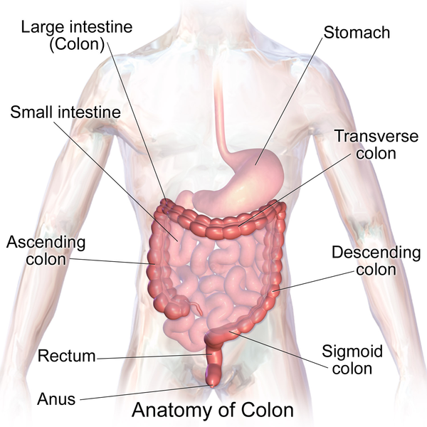
The Large Intestine (Image Source). Here, the difference pieces of the large intestine have been labeled. The first piece is the ascending colon, which is connected to the small intestine and carries food up. Then comes the transverse colon, which carries food across (from the left side of the picture to the right side of the picture), followed by the descending colon and the sigmoid (“s-shaped”) colon, which move processed food down into the rectum.
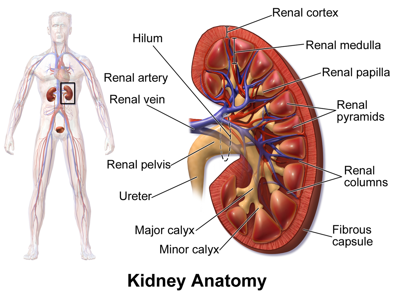
The Kidneys (Image Source). The inset on the left shows the two kidneys relative to the whole body. If you look closely you can see the ureters leading down from the kidneys to the bladder. The kidney has many sub-components to filter the blood and produce urine. Food for thought (or, water for thought): When you drink a glass of water, your intestines absorb that water into your bloodstream. Then that glass of water gets filtered back out of your bloodstream by the kidneys – i.e., it’s turned into urine. So all of the water that you consume has an “intermediate stage” where it is part of your blood.
The end. Congrats – you’ve finished the intro to abdominal anatomy!
Want to be the first to hear about my articles bridging healthcare, artificial intelligence, and business—and get a free list of my favorite health AI resources? Sign up here.
