

It is useful to be familiar with basic chest anatomy when analyzing chest x-ray or chest CT images. First I’ll do an intro to the different organs and structures in the chest, and then I’ll go over some images showing their locations.
Organs & Structures of the Chest
- Heart. The heart is a four-chambered organ that pumps deoxygenated blood through the lungs and pumps oxygenated blood through the body.
- Atria (right atrium and left atrium): the small, upper chambers of the heart
- Ventricles (right ventricle and left ventricle): the large, lower chambers of the heart
- Superior vena cava and inferior vena cava: big veins that carry deoxygenated blood into the heart. The superior vena cava comes down from above and the inferior vena cava comes up from beneath.
- Aorta: the big artery that carries oxygenated blood to the body
- Lungs: organs that add oxygen to the blood and remove carbon dioxide from the blood. (They are not hollow; they have a spongy consistency to enable massive surface area. The total surface area of your lungs is about the same as the surface area of one side of a tennis court.)
- Right upper lobe & left upper lobe (also called the superior lobes)
- Right middle lobe & lingula: the right lung has a middle lobe, and the left lung has what’s called the “lingula” (“tongue”) which is a tiny version of the middle lobe
- Right lower lobe & left lower lobe (also called the inferior lobes)
- Larynx: the voice box
- Trachea: the windpipe
- Bronchus: the trachea splits into two tubes; these tubes are called bronchi. (It splits into many more smaller tubes after the bronchi. These tiny tubes carry air through the spongy lungs.)
- Diaphragm: a thin dome-shaped muscle that helps with breathing. It is located between the chest and the abdomen.
- Pleura: the sacs that surround the lungs. The “pleural cavity” is the inside of the sacs, and is the space where the lungs sit.
- Mediastinum: the heart and great vessels. The mediastinum does NOT include the lungs. You can think of the mediastinum as the “middle” of the chest (“media”). (As my anatomy professor used to say, if you learn nothing about chest anatomy, learn that “the lungs are not in the mediastinum. The lungs are not in the mediastinum…”) This word is pronounced “media – STY – num” (“sty” rhymes with “fly”)
Pictures of the Chest
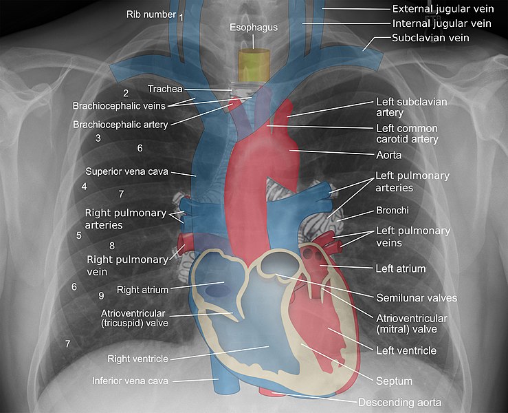
Diagram of the Mediastinum (Image Source.) In the diagram above, the blue and red colors indicate the heart and great vessels. The blue parts carry deoxygenated blood and the red parts carry oxygenated blood. The red tubes and the blue tubes on the top are huge blood vessels. You can see the red aortic arch with three other blood vessels coming off of the top. It is hard to see the windpipe (trachea) in this picture because it’s behind all of the blood vessels and the heart. You can see the four chambers of the heart: the right atrium, left atrium, right ventricle, and left ventricle.
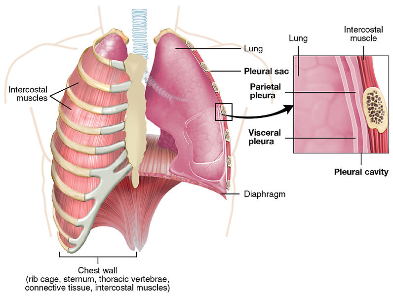
Diagram of the Lungs (Image Source.) In the diagram above on the left, you can see the ribs and the “intercostal muscles” (muscles between the ribs), which are completely covering up the whole lung, except for the very tip of the lung (it’s drawn in pink; you can see it poking out at the top). On the right, the ribs and the intercostal muscles have been “removed” so you can see the entire lung. The outside of the lung is covered in the pleural sac, also known as the “pleura.” Below the lungs you can see the diaphragm, a big dome-shaped muscle. When you get the hiccups, it’s because your diaphragm is spasming.
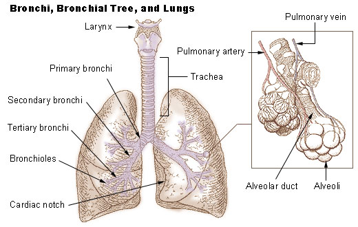
Another Diagram of the Lungs (Image Source). The very top of this image is your larynx, where you have your voice box. If you speak and place your hand over your throat you can feel the approximate location of your larynx. Below the larynx is the windpipe (trachea), which splits into bronchi. The bronchi split repeatedly until they become bronchioles, which are about one millimeter or less in diameter. Actual exchange of oxygen and carbon dioxide happens in minuscule sacs called “alveoli”; these sacs are the final destination of the air you breathe in.
Links to Additional Diagrams
- Cartoon of lungs and heart
- Another cartoon of lungs and heart (encased in ribs)
- Cartoon of the lobes of the lung
- Detailed cartoon of the heart, lungs, and great vessels
I am linking to these diagrams instead of pasting them here because they are copyrighted images.
That’s all folks! Stay tuned for future posts about chest x-ray and chest CT interpretation!
Want to be the first to hear about my articles bridging healthcare, artificial intelligence, and business—and get a free list of my favorite health AI resources? Sign up here.

Comments are closed.